Skeletons are laid out in the laboratory
With the analysis of non-cremated, articulated human remains, the skeleton is laid out in the laboratory and arranged in the correct anatomical position.
The remains are photographed and recorded using standards and recording sheets published by Brickley & McKinley (2004). The preservation, completeness and condition of the bones are recorded using IfA guidelines (ibid). The osteoarchaeologist will then examine the skeleton to build a biological profile of the individual.
Analyses can reveal:
- Age-at-death (using sternal rib end analysis, molar wear patterns, clavicular fusion, pelvic traits, epiphyseal fusion rates in neonatal and juvenile remains)
- Sex (morphological traits on the pelvis and cranium; lengths of femora & radii)
- Ancestry
- Palaeopathology such as infectious diseases (syphilis, tuberculosis), degenerative joint diseases, metabolic diseases such as scurvy, rickets, iron deficiency anaemia, genetic diseases
- Genetic conditions such as cleft palate
- Dental Pathology – gum disease, cavities, calculus (solidified dental plaque), abscesses
- Trauma – fractures (healed/non-healed), cut-marks - signs of violence
- Metric analyses such as stature and body mass
- Musculo-skeletal markers – can provide a tentative suggestion of occupational activities
- The presence of cranial and post-cranial non-metric traits can provide information about family traits and ethnic origins e.g. metopic sutures on the cranium
In order to build a full biographical profile of the individual, other factors are also considered, such as:
- Overall plan, shape and depth of the grave (if applicable)
- Quantity & type of grave goods (if any) – can be gender-specific
- Position of the body in the grave
- Orientation of the grave (N-S, E-W)
- Was it a single or multiple burial? If it’s a multiple burial, are the individuals related?
- Taphonomy and the burial environment are also considered e.g. soil pH, root damage
If necessary, small samples of human bone will be sent for radiocarbon dating and isotopic analysis. Collagen-rich anatomical elements include phalanges and femora as well as carpals and tarsals.
Benefit to the developer
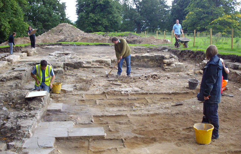
A recent archaeological excavation was undertaken by Wardell Armstrong Archaeology at Ettington Road, Warwickshire. A single, poorly-preserved, articulated inhumation was discovered at the base of one pit.
The skeleton was carefully excavated and recorded.
Having experienced staff on-site was a huge benefit, as the skeleton could be recorded and lifted quickly, which primarily aided the long-term preservation and curation of the remains but also ensured the effective completion of the project.